The knee joint
 The knee is the largest joint in the body and essential to many of our daily activities including standing, walking, running and sitting. We wouldn’t get far without it! Unfortunately, it’s also prone to injury, causing millions of Americans to visit their doctors with knee complaints each year.
Anatomy
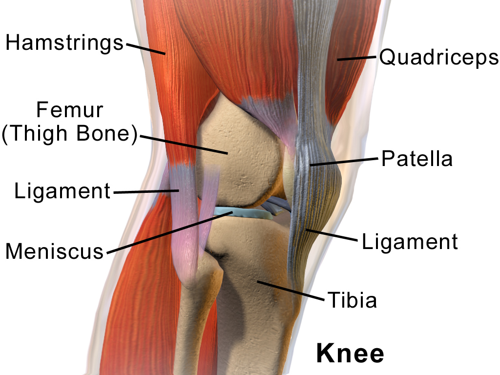
The knee is a synovial hinge joint made up of three bones: the femur (thigh), the tibia (shin) and the patella (kneecap). It’s surrounded and mobilized by several groups of large, powerful leg muscles; held together and supported by strong ligaments, both outside and inside the joint; cushioned by cartilage; and lubricated by synovial fluid.
Movement
The knee is able to move in four directions. Its main movements are flexion (bending) and extension (straightening). It’s also able to rotate in and out a small amount when it’s flexed. When the knee is straight all of its rotation happens further up the kinetic chain at the hip joint. The knee’s movement is articulated by several groups of strong leg muscles:
Knee extensors

The quadriceps femoris
The quadriceps femoris are the long front thigh muscles. They originate at the hip and upper thigh, and insert at the shin bone below the knee. Their job is to extend the leg straight and help track the patella in place. If their strength is off balance (one quadricep is stronger or weaker that the others) they can pull the patella off track and cause pressure to load unevenly on the joint. Also, if they become too tight, they can prevent the knee from fully flexing.
The quads are made up of four different muscles:
- Rectus femoris: the most superficial of the quadriceps muscles. It extends the leg and is the only quad muscle to attach to the pelvis, so it also acts as a hip flexor.
- Vastus lateralis: located on the outside of the thigh, often stronger than the other vastus muscles.
- Vastus intermedius: located at the center of the thigh, beneath the rectus femoris.
- Vastus medialis: located on the inside of the thigh, this muscle plays an important part in stabilizing the kneecap. It’s often found to be weaker than the other vastus muscles when knee pain is present. This is in part due to it only fully engaging during the last 10-15 degrees of extension.
Knee flexors
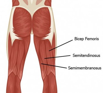 The hamstrings
Several muscles are involved in knee flexion: the hamstrings, gracilis, sartorius and popliteus.
The largest group are the hamstrings, long muscles that run down the back of the thigh. They help bend the knee and stabilize it by preventing torsion from too much twisting at the joint. They’re antagonists (opposing) to the quadriceps, and if they’re too tight they can prevent the quads from fully straightening the leg. Tight hamstrings can also negatively impact the knee cartilage.
There are three hamstrings that attach from the buttock bones to the back of the shins:
- Semitendinosus: flexes the knee and inwardly rotates the lower leg when the knee is bent.
- Semimembranosus: same actions as the semitendinosus.
- Biceps femoris: flexes the knee and outwardly rotates the lower leg when the knee is bent.
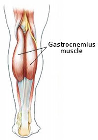
The calves
The gastrocnemius is the largest and most superficial calf muscle at the back of the shin. It helps flex the back of the knee and plantar flexes the ankle (points the foot). It’s a strong and important walking muscle. Every step you take is aided by contracting gastrocnemius muscles, and because of this it gets tight easily. This can lead to reduced range of motion at the ankle and the back of the knee, both of which can affect your walking gait and negatively impact the knee.
Honorary mention
The Iliotibial tract
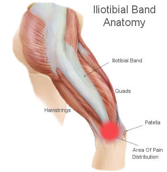 The iliotibial tract is a long, deep band of fascia (connective tissue) that runs from the outer hip down to the outer shin. It supports the knee in both flexion and extension, and is therefore in constant use when we’re walking or running. Because of its high activity levels, it’s prone to tightness and sometimes strain. Iliotobial band syndrome is a condition that occurs due to overuse or irritation of the band rubbing up against the outer knee bones, and can cause pain and swelling at the outer knee. Strengthening the hip muscles and stretching the outer thigh can be a great help, along with wearing proper footwear, especially for running.
Connect
For more yoga articles, updates, classes and workshops, sign up for my newsletter at the top of the page or like on Facebook at Ann West :: Iyengar Yoga. You can contact me directly by email or call (858) 224-2484.
© 2017 by Ann West. All rights reserved.
Leave a Reply